
Ultrasound Services
MD Ultrasound is offering ultrasound diagnostic services to physicians and their patients. Our professional sonographers can travel to physicians’ offices or any other health care facility, as well as provide services at our primary location in Burbank, CA.
Test results are interpreted by certified radiologists and/or cardiologists. The final report of patient is conveniently accessible to providers online from any secure computer. We contact the patient within a business day for appointments and allow access to patients’ reports and images online. Referring physicians will be able to download the reports and images of their patients any time.
Give us a call at 747-229-9000 or book an appointment
Skin Ultrasound
High resolution (greater than 15 Mhz) ultrasound imaging is increasingly being used in the evaluation of various medical conditions of skin. Ultrasound is the only imaging modality useful in the evaluation of skin lesions that are too small to be seen on computer tomography (CT) or magnetic resonance imaging (MRI). Noninvasive sonographic investigation is expected to determine the size, contour and structure of skin lesions.
- Benign and malignant neoplasms (melanoma, basal cell carcinoma, hemangioma, fibroma, seborrheic wart)
- Inflammatory and infectious diseases (systemic scleroderma, lipodermatosclerosis, psoriasis)
Evaluation of lymphedema of the limbs, wound healing, scars, nail lesions
Evaluation of lumps, bumps
Ultrasound supports clinical diagnosis and local staging of some tumors (epithelial), helping to detect tumor borders for complete excision. Use of Doppler during the exam shows blood flow around the area of question or vascular structures.
Cosmetic dermatology is another field of diagnostic ultrasound implementation. In the era of cosmetic fillers and aesthetic treatment procedures ultrasound is used to assess skin response to laser, mesotherapy, photodynamic therapy as well as in the event of different complications.
Neuromuscular Ultrasound
High resolution ultrasonography allows the clinician to examine peripheral nerves such as median, ulnar, radial, sciatic, fibular, and obtain detailed structural images of them. Dynamic evaluation of nerves in different angles during an extremity movement detects the changes of echostructure in the real time. Focal neuropathies, motor neuron disease, polyneuropathies, muscular dystrophies and myopathies are common neurological pathologies detected by high resolution nerve and muscle ultrasound.
Navigational capabilities of ultrasound in the Interventional and Regenerative medicine has approved by medical and surgical communities. Whether it’s come to Myofascial Pain Syndrome, Trigger Point localization, PRP injection, Hydrodissection of pinched nerve root, Radiofrequency or Cryoablation of peripheral nerves ultrasound guidance has been approved as an essential tool for a specialist.
Nerve ultrasound is an anatomical evaluation of the peripheral nerves whereas electrophysiological studies of the nerves such as NCV and EMG are considered physiological. Magnetic Resonance Imaging (MRI) allows detailed imaging of peripheral nerves and their pathology. However, MRI is costly and time-consuming method of nerve mapping that only provides static imaging. Nerve ultrasound is an alternative imaging method for localization and examination of peripheral nerves. Nowadays ultrasound guided nerve blocks are used before and during physical therapy. Therapeutic courses of ROM improvement are better tolerated after ultrasound-guided therapeutic pain injections and nerve blocks. With pain relief the patient will get improved motility of extremity. That means adequate physiotherapy and limb-strengthening exercises can be carried out.

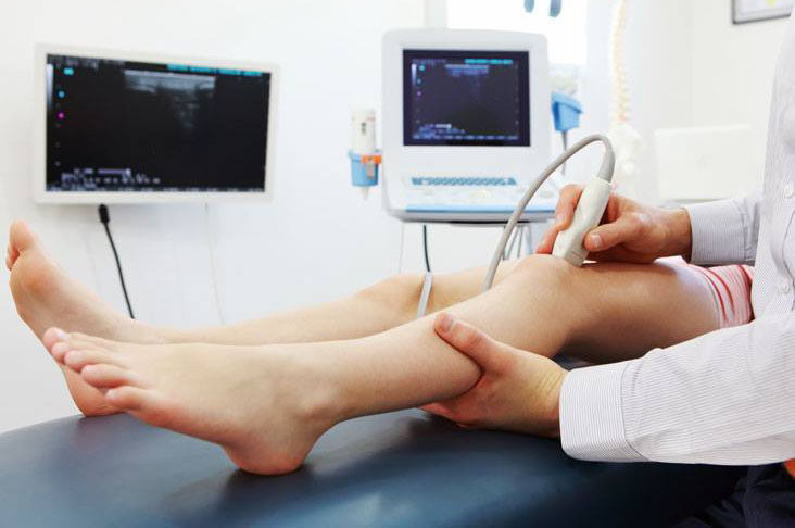
Musculoskeletal Ultrasound (MSK)
Musculoskeletal ultrasound (MSK) is very useful noninvasive examination of muscles, tendons, ligaments and bones. It may be prescribed for various problems of neck and cervical spine, shoulder, knee, ankle and other joints. Inflammation, osteoarthritis, tendinopathies, entrapment neuropathy such as carpal tunnel syndrome or piriformis syndrome – this is the incomplete list of pathological conditions.
Modern ultrasound machines equipped with high resolution transducers allow to see anatomical structures in more detail. Live ultrasound testing (examination while body parts are in motion) is more valuable for the physician than stationary images as in MRI/CT.
Ultrasound examination of the musculoskeletal system is typically used to diagnose:
- Ligament tears or sprains
Muscle tears
Tendon tears or inflammation, such as rotator cuff in the shoulder
Inflammation of joints and fluid collection (effusions)
Benign or malignant tumors of soft tissue
Arterial, Venous Ultrasound
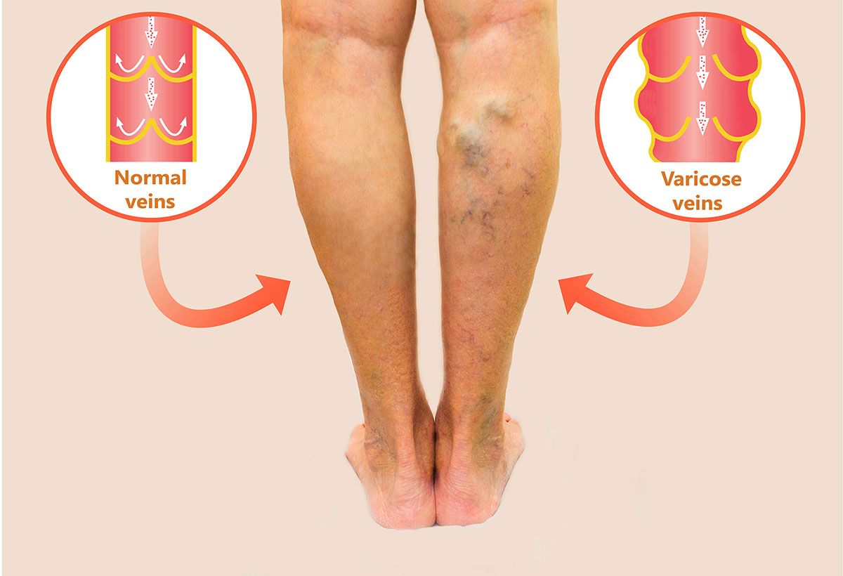
Arterial and Venous studies of lower extremity – doctors order these tests in: pain, cramping, tingling and swelling of lower legs; varicose dilatation of veins, skin thickening and darkening. Dupplex sonography is a first line diagnostic and confirmative test for deep venous thrombosis (DVT). This pathological condition is very dangerous and is not rare after surgeries of legs and pelvic organs such as total knee replacements or radical prostatectomy for prostate cancer. If not recognized by Doppler ultrasound and prevented effectively, DVT of legs may cause venous thrombi to break off to form pulmonary emboli.
Swollen legs, pain and numbness during a physical activity, skin discoloration or thickening in lower legs – these are common medical conditions. Duplex examination of arteries and veins is of medical necessity to find an atherosclerotic plaquing and stenosis of arteries as well as venous insufficiency or reflux.

Ultrasound Guidance
We provide ultrasound imaging and guidance to many interventional procedures. Our experienced sonographers can help in different procedures done by ultrasound navigation. Many vascular surgeons, pain physicians, neurologists, orthopedists, sport medicine physicians need not only ultrasound imaging before and after the procedures but also during the intervention itself. Ultrasound guidance is essential in many interventional procedures on veins, arteries, joints, nerves and soft tissues.
- Chronic and acute pain management
- Musculoskeletal, joint and soft tissue injections. Accurate needle placement.
- Peripheral nerve blocks before and after the surgery.
- Venous sclerotherapy, endovenous laser treatment (EVLT) of varicose veins, arterial revascularization in limb ischemia and arterial insufficiency
Urology
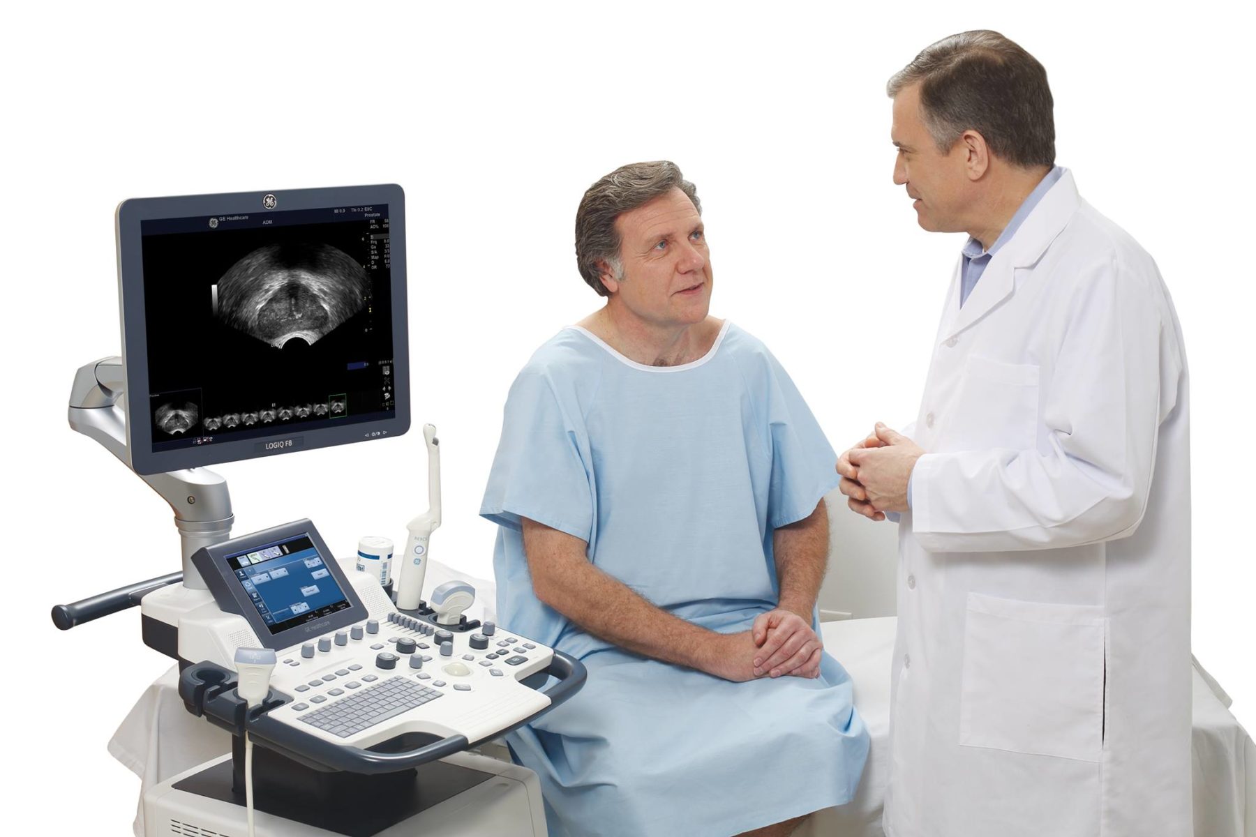
Ultrasound examination is very valuable and sometimes is first line diagnostic tool in urological problems. The ultrasound is very effective in diagnosing urinary stone disease, cysts and tumors.
Prostate enlargement, prostatitis, and mass lesions in the urinary bladder are easily revealed by ultrasound examination. In suspicious prostate cancers, ultrasound is done by transrectal root (TRUS). Testicles are examined by the ultrasound for different conditions: torsion, varicocele, hydrocele, male infertility. Doppler examination of male genitalia has its specific role in diagnostics of impotence and sexual dysfunctions.
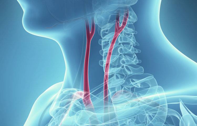
Vascular Ultrasound
Carotid arteries carry a blood from heart to the brain. Atherosclerotic thickening of intimal arterial wall, calcified plaque formation in these arteries may block the blood flow and cause brain stroke.
The Abdominal aorta is the biggest artery of our body carrying blood from the heart to inner organs and lower extremities. Ultrasound of abdominal aorta is the recommended screening test for those adults who is 65 and older and who has ever used tobacco products.
TCD (transcranial Doppler) mostly prescribed by neurologists for different medical conditions of our brain as well as for stroke prevention.
Gynecology
Gynecology: female pelvic, uterus and ovaries might be seen by using both transabdominal and transvaginal probes. The transabdominal assessment usually provides overview of the pelvis, whereas the transvaginal ultrasound creates more precise and detailed images, especially in obese women.


Lung Ultrasound
NEW SERVICE from Imaging Center – Lung Ultrasound screening for pneumonia and pleural effusion. In the current situation with virus spread (COVID-19 and others), it’s preferable to have a simple non-invasive diagnostic assessment of the lung which will reveal potential pneumonia. Rapid test result is essential for making the right decision and hospitalize the patient on time, if needed.
Other Ultrasound Services
Abdominal ultrasound test is done for various medical problems of the liver, gallbladder, kidneys, spleen, abdominal aorta. Fatty liver, gallstones, kidney stones, spleen enlargement, fluid collection, various types of tumors are identified.
Thyroid ultrasounds are a useful tool to find possible nodes, cysts, tumors in thyroid gland. Some medical conditions may be related to thyroid function, such as heart arrhythmia, change of weight, depression etc.
Echocardiography ordered by physician in certain heart conditions such as: chest pain, shortness of breath, hypertension disease, mitral valve prolapse, myocardial infarction, brain ischemia etc.
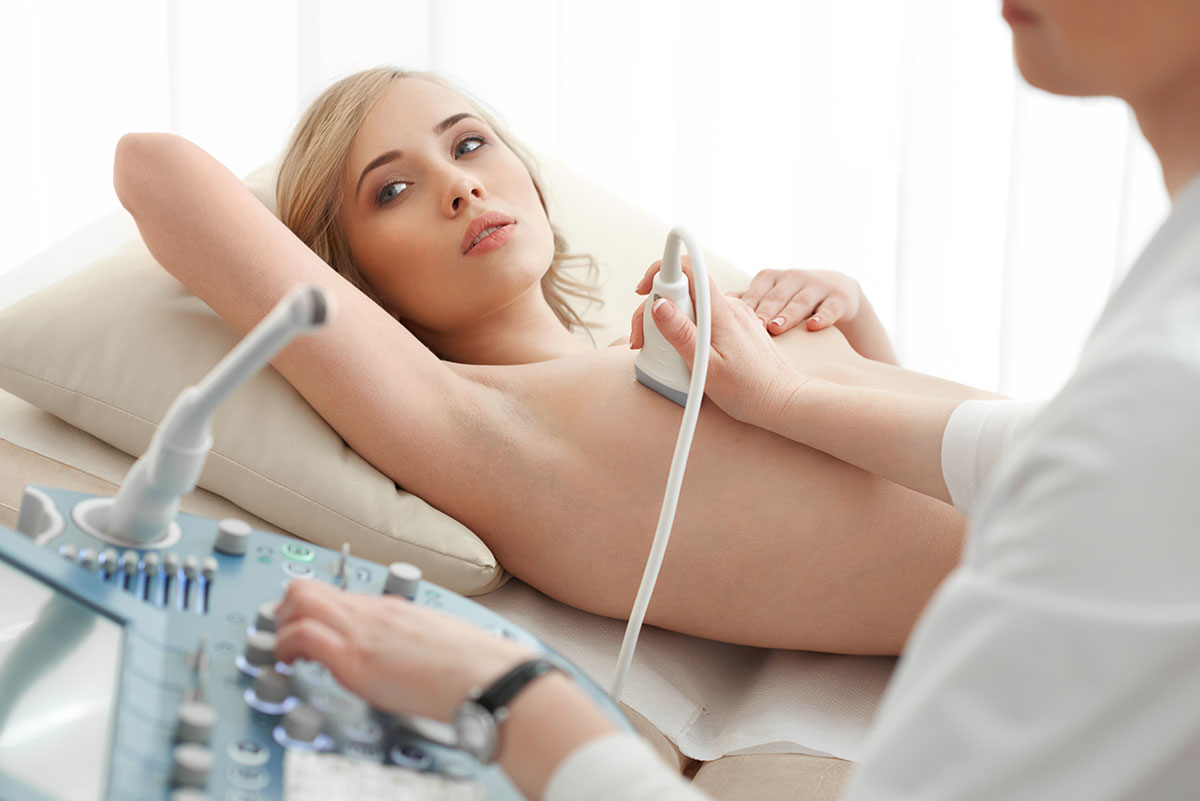
Breast ultrasound is safe, painless and does not expose any radiation to the body.
 Call us